Anatomy of inguinal canal:
Just above the medial (situated in or extending toward the middle) half of inguinal ligament in the anterior (Placed before or in front) abdominal wall, there is a passage that allows spermatic cord in men and round ligament in women to pass. In adults, it is about 3.75 cm long directed downwards and medially running from the deep inguinal ring to superficial inguinal ring. A triangular opening in the aponeurosis (sheet like tendinous expansion) of external oblique muscle 1.25 cm above the pubic tubercle is called superficial inguinal ring. Deep inguinal ring is U-shaped and lies 1.25 cm above the point on inguinal ligament midway between the anterior superior iliac spine and pubic symphesis (fibrocartilaginous fusion between pubic bones).
Inguinal canal is very important part of the body transmitting the spermatic cord, the ilioinguinal nerve and genital branch of genitofemoral nerve (a filamentous band of nervous tissue arising from lumber region) in men. In women, round ligament is found in the place of spermatic cord (Figure 1).
Inguinal canal is very important part of the body transmitting the spermatic cord, the ilioinguinal nerve and genital branch of genitofemoral nerve (a filamentous band of nervous tissue arising from lumber region) in men. In women, round ligament is found in the place of spermatic cord (Figure 1).
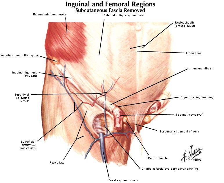
Figure 1: Anatomy of the inguinal canal.
|
Anatomy of femoral canal:
Femoral canal is a vertical passage of 1.25 cm long and 1.25 cm wide that travels from femoral ring above to the saphenous opening below and occupies the most medial (inside) compartment of the femoral sheath (A band of fibrous tissue located in the upper thigh, through which pass the main femoral artery, vein, and lymph vessels). The contents of femoral canal include lymphatic vessels, lymph nodes and fat (Figure 2).

Figure 2: Anatomy of femoral canal.
|
